by Arnie Schoenberg
version: 13 July 2023
Figure 3.5 human female skeleton, red lines point to individual bones, blue lines point to groups of bones by Mariana Ruiz Villarreal, 2007 (Public Domain)
Osteology is the study of bones. Osteology is important to studying human variation, and primatology. Paleoanthropology relies on osteology because most fossils come from bones. Forensic anthropology uses osteology to solve crimes.
Like most other physical traits, the bones we see are a consequence of genes and environment. There is nothing particularly profound about bones compared to other biological systems, but their durability makes them special for anthropology because they are the main source of data for paleoanthropologists, important to archaeology, and before DNA testing, they were important to the study of human variation.
We tend to think of bones as dead, dry, and brittle, and when you leave them out in the sun for a few years they do get old. Their hardness comes from a calcium-based crystal structure. The molecules interconnect like columns of Lego blocks.

Figure 3.1 Hydroxyapatite molecule, by J. Kirkham © 2007
In a biology class you tend to think of a bone as a living organ, like your heart or your lungs, but in anthropology we are used to looking at dead bones, outside of the body, when they are just shells of the functions they had when they supported living organisms.
Figure 3.2 * Bone Growth by rozwój kos«ci (public domain)

Figure 3.3 Periosteum and Endosteum by OpenStax, College Anatomy & Physiology, 6.3 * Bone Structure, 2018 (CC-BY-4.0)
Genetics determines most of what your bones look like. For example, your 23rd chromosomes determine several shapes that are commonly used to say whether someone looks male or female, and forensic anthropologists use these differences to identify the sex of a skeleton.

Figure 3.4 to adapt to reproductive fitness, the female pelvis is lighter, wider, shallower, and has a broader angle between the pubic bones than the male pelvis. by OpenStax, College Biology, 38.1 * Types of Skeletal Systems, 2018 (CC-BY-4.0)
But like the rest of your body, the environment also effects your physical structures. The muscle attachments on your bones suggest your activities during your life, and stress, i.e. malnutrition, can be read in cross-sections of your teeth like tree rings.
It's important that we have a basic shared vocabulary so that we can compare humans to other vertebrates, to evaluate fossils, and to understand several aspects of human variation.
LEARN THE BONES OF THE HUMAN SKELETON BELOW:
Figure 3.5 basic human female skeleton, red lines point to individual bones, blue lines point to groups of bones by Mariana Ruiz Villarreal, 2007 (Public Domain)
If you want to memorize these bones try clicking the blank one, print a few copies out, and practice writing the names of the bones. Check your spelling and try to learn the scientific names.
| sit bone | ischium |
| tail bone | coccyx |
(adapted from George Claypoole http://hes.ucfsd.org/gclaypo/skelweb/skel03.html)
here's an online practice quiz with more detail then you need for this class, but good to know if you're going on to study anything health related.
skim animal skeletons
SKIM PRIMATE SKELETONS
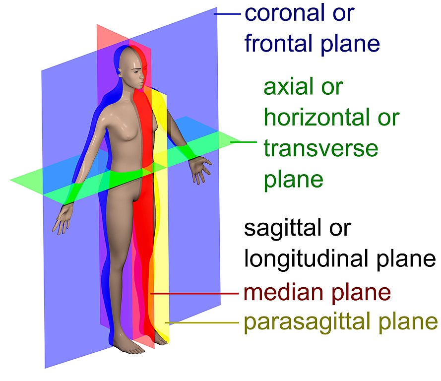
Scientists have agreed on naming conventions to refer to points on the body, such directions and planes. These correspond to several of the bones and sutures in the skull.

Figure 3.6 Human Anatomy Planes by David Richfield and Mikael HŠggstršm, M.D., via Wikimedia Commons (CC BY-SA 4.0)
Vocabulary
Imagination Questions

Figure 3.6 hah-ah-ah-hahhahaha by Allison Zai from Skeletons © 2018 (permission pending)
previous section: 2 intro to biology
problems (mistakes, bad links, etc.)